Anesthesiologist & Artist
The photographs of Alfred Feingold, M.D.
|
|
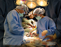
PHYSICIAN AND PHYSICIAN ASSISTANT: Cardiothoracic surgeon Lawrence Dacey, M.D. (left), and
physician assistant Ryan Hafner are replacing a heart valve. The foreground image was taken in
the middle of the procedure, while the surgeon was using a suction catheter to siphon blood away
from the incision. The background image, showing only the hands, was taken later, while the
incision was being closed. Dacey has been on the faculty since 1993; he did some of his training
at DHMC and also holds a master's degree from Dartmouth's evaluative clinical sciences program.
|
|
One of the hardest things to do is to get
the right scale. If you back off to show
the lights and the faces, then you lose
the detail in the hands. And if you move
in to show the hands, then you miss the
concentration of the surgeons. This
overlay allows me to do both. ORs are
a very difficult area to photograph
because in a lot of work, the meaning
comes from the face—from the nose,
from the lips. When you take surgical
pictures, you lose this important
dimension of human meaning. All I have
is the eyes, the hands, and the body
posture. It's not easy to tell a story
with the nose and the lips covered.
|
|
|
|
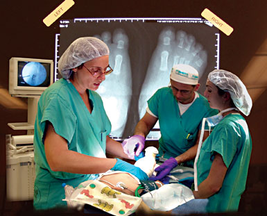
UNMASKED: Orthopaedic surgeon Kathleen Moen, M.D. (left), is working here with resident Jorge Brito, M.D. (center),
and Jessica Pelow, a fourth-year medical student at the University of Buffalo who was doing a surgical rotation at
DHMC. They are not wearing masks because they've just finished a procedure; they're now putting a cast on after
having removed an extra toe from a child's foot. A larger-than-life-size x-ray appears to be taped to the wall behind
them. And the overhead fluorescent lights, angled in on both sides of the frame, resemble rays of sunlight.
|
| |
|
I had an earlier image of
Moen with a mask on.
She looked just like
anyone else. So when I
saw this one with her
mask off—and I saw it
came out well—I knew I
had to work with this
image. The fluoroscope
machine [on the left]
shows what the operation
was. It shows that there
are small pins or nails
that they put in the
child's foot—that's the
fixation device used
in this operation.
|
|
|
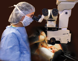
INTRICATE MOVES: Eye surgeon Susan Pepin, M.D., is
using microsurgical techniques to perform a cataract
extraction. After she removes the cataract—a lens that
has become cloudy—she'll replace it with a synthetic lens.
|
| |
|
The interesting thing about eye surgery is that the
surgeon is almost motionless, as compared to other
operations where the body and arms are moving
around. Ophthalmological surgeons are almost
frozen, with their hands making intricate, small
motions. I got a picture of this surgeon looking
through the operative microscope, and then
dropped behind it a picture of her hands and the
eye. With ophthalmologic surgery, the light is so
bright that I don't need any other light. In fact,
I have to stop down my camera to get a decent
picture—otherwise the light wipes out the image.
|
|
|
|
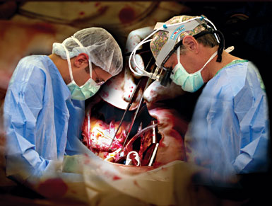
FROM THE HEART: Resident S. Scott Lollis, M.D. (left), is helping cardiothoracic surgeon William Nugent, M.D., do a
coronary artery bypass. Nugent has been on the faculty since 1983; Lollis is a 1998 graduate of Dartmouth College.
|
| |
|
Here the foreground is
being enveloped by the
background. And notice
the attention, the
concentration, the body
posture of the surgeon
and the resident. You can
tell this is a coronary
bypass because these
are the kind of catheters
they put in the heart
when they do a bypass.
And the patient's on a
heart-lung pump, too—
you can see the spiral
tube that sucks blood in
and out of the heart.
|
Previous Page | Next Page
Back to Main Article