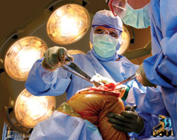
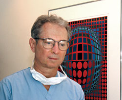
Anesthesiologist & Artist
The photographs of Alfred Feingold, M.D.
|
|
|
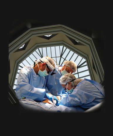
SENSE OF ACTION: The skylight above DHMC's main rotunda has been applied as a frame around these three individuals who are using a fiber-optic device to examine valves in a leg vein. From left to right are resident Michael Alvarado, M.D., technician Michael Poulen, and vascular surgeon Eva Rzucidlo, M.D. Alvarado is a 1998 graduate of DMS, and Rzucidlo completed a fellowship in vascular surgery at DHMC in 2002 and then joined the faculty. |
|
This works because she's paused, she's in sharp focus. But the surgeon on the left, his hand is moving—you get a sense of action, of motion. That is not easily achieved. This was an operation on the whole leg, from the ankle up to the hip. I was trying to come up with an image to show the surgery, but I just couldn't get the long incision to fit. This is one of the images I came back to several times— and all of a sudden I looked at it again and remembered the picture of the rotunda and said, "Wow, the rotunda works." When we were walking down the mall, I recall saying, "Let's stop—I want to get a picture of this rotunda." And note that the earring identifies the surgeon on the right as a woman. Whenever I can I show jewelry—in fact, I will sharpen the highlights— so it's clear it's a woman. |
|
|
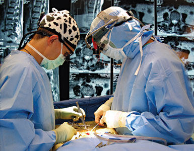
RADIOLOGY AND RETINAS: Orthopaedic surgeon William Abdu, M.D. (right), and resident Eric Marsh, M.D., are performing a type of back surgery here—a laminotomy and intervertebral disc excision, meaning removal of the herniated portion of a disc through the lamina, the bony covering over the spinal canal. Feingold used the patient's x-rays and MRI scans to wallpaper the background of the image, contrasting the insight they offer with the insight offered through the surgeons' eyes. Abdu is an alumnus of DHMC's residency program ('85-90) and also earned a master's degree from Dartmouth's evaluative clinical sciences program in 2001. |
|
X-rays make great backgrounds. This image tells the story of this operation—the fact that the diagnosis was made by x-rays. The eyes of the x-ray machine allow the surgeons to use their own eyes. There are two different types of sight—the x-ray sight and the surgeon sight. Here, you see the x-rays in black and white, and then you have the surgeons in color. |
|