Vital Signs:
DHMC radiology
aims at a world
without any film
In a dimly lit radiology reading room at DHMC, several radiologists flip through 80 digital "slices"-cross-sectional images of a patient's liver-on a computer screen. They are using a state-of-the-art picture archiving and communication system (PACS). With just a few clicks of a mouse, a doctor inverts an image for better contrast, zooms in to get a closer look at a tumor, then uses an electronic measuring tool to figure its exact size.

|
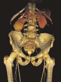
| This 3D digital image of a hip is a product of new filmless imaging technology. |
Since PACS was installed at DHMC in February 2003, radiology has been moving quickly toward an almost totally filmless environment. PACS has "made life a lot easier" for technologists, radiologists, clinicians, and patients, says Dr. Peter Spiegel, chair of radiology. With the new system, technologists can produce images-CTs, MRIs, or xrays- much faster than when they had to develop film. And it's more efficient for radiologists, too, since they don't have to spend time handling film.
"A tremendous advantage of PACS is its speed and the facility it gives us," says Spiegel. "If you want to know if a patient has had an echocardiogram, you can look into the physician's notes or at the lab data and keep the corresponding images up on a second screen, moving back and forth." A CT or MRI case can include more than 100 images. With PACS, it's much easier to process that kind of volume.
Referring clinicians like the system, too, since they have immediate access to images on their laptops. "If you get calls in the middle of the night," says Dr. Clifford Eskey, "you can access full-fidelity images . . . at home. It's very reliable."
Patients also benefit, since they don't have to wait as long for images to be processed; usually, a patient can meet with the referring clinician 30 minutes after an image was taken. "The patients polled have been very impressed . . . with the technology and the ability to see their images manipulated before their eyes. It instills a level of confidence," says Ned Turpin, director of clinical operations and ancillary systems.
Kneecap: If a patient is suffering from knee arthritis, for example, an orthopaedist can display an image of the kneecap and then zoom in to show details of bone spurs and other early signs of osteoarthritis. The PACS project was divided into three phases, says Monte Clinton, administrative director of radiology. Surgical radiology came first. In 2002, DHMC purchased six radiographic rooms and started archiving digital CT, MRI, and ultrasound images. Then 22 42-inch, flat-panel, plasma monitors were installed in the ORs. The second phase, diagnostic radiology, was completed in 2003. The third phase, nearly finished, involves nuclear medicine and interventional radiology. Mammograms are being converted to digital form gradually through the whole project.
To create images, radiology technicians now use a variety of high-tech devices. A 16-row Lightspeed scanner produces intricate, three-dimensional, color angiographic images. DHMC also recently purchased an automated digital radiographic room, one of only three in the world. It has a machine that operates as an upright detector for chest exams and as a table detector. "This is a very unique concept, and it makes the room more versatile and less costly. The detector is moved by a robotic system," says Clinton.
The department's 20 radiologists, 60 technologists, 8 nurses, and 82 support staff handle 300 to 400 patients on an average weekday. PACS has led to "large increases in productivity," says Clinton. The department did about 200,000 exams in fiscal year 2003 and expects to do 218,000 in 2004, says Clinton- a nearly 10% increase.
PACS has also brought substantial financial savings and environmental benefits. "DHMC no longer has to [buy] $600,000 worth of film each year," says Clinton, "or dispose of one million gallons of water each year to process films."
Matthew C. Wiencke
If you would like to offer any feedback about this article, we would welcome getting your comments at DartMed@Dartmouth.edu.
