
Biochemist explores mysteries of mitosis and motor proteins
Duane Compton, Ph.D., an associate professor of biochemistry, is deeply involved in the study of a life process that is as fundamental as it is murky.
Prior to cell division, the last phase of the cell cycle, the newly replicated chromosomes produced in the synthesis (S) phase are partitioned equally into separate parts of the cell. The cell then divides in such a way that each daughter cell has an identical set of chromosomes.
Spindle: During that process, the cell first constructs and then disassembles an elaborate structure in its interior called a spindle; this structure exists only temporarily, for the sole purpose of capturing, aligning, separating, and strategically locating the two sets of chromosomes. The spindle has a distinctive appearance when viewed microscopically and gets its name from its similarity to a tapered pin, or spindle, used in spinning.
At the start of mitosis, subcellular structures called centrosomes migrate to opposite sides of the nucleus and begin assembling long tentacles of microtubules, which are organized by a group of accessory proteins. Identifying those important accessory proteins and elucidating their precise function is what occupies Compton and the corps of eager graduate students and postdoctoral fellows in his lab.
He describes the process of spindle-formation "as similar to picking a bouquet of flowers, which are held together in one hand. The hand that holds the stems represents a structure that is called the spindle pole, which evolved from the centrosome, the stems represent the microtubules, and the blossoms represent the chromosomes."
The organization of the "hand," or spindle pole, involves the contributions of specific proteins that are of interest to Compton's lab. The short ends of the stems/microtubules are anchored to the interior of the cell wall, while the nuclear membrane disintegrates so the long ends can reach the chromosomes.
After the DNA strands have been duplicated in the S-phase of the cell cycle, they remain attached to each other in a constricted region called the centromere, forming an X-shaped structure called sister chromatids. The centromere on each chromosome has a specialized structure called the kinetochore, which is the precise target for its specific microtubule.
Strands: When each microtubule is attached to its kinetochore, the stage is set to pull the sister chromatids apart and separate the twin strands of DNA. Some theorists have imagined the microtubules as cables that act in concert to pull the strands apart and tow them into the separate regions of the cell destined to become the daughter cells.
But no little "elves" pulling on the ends of the cables have ever been identified, so this tug-ofwar analogy was scrapped.
Approximately 22 microtubules in a human cell must grow from each spindle pole and attach to each kinetochore, forming a shape somewhat like a football —the spindle.
|
|
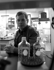
Duane Compton is investigating aspects of cellular division
—work that may lead to new cancer therapies.
|
For reasons that are not entirely clear, the chromatid sisters are first aligned together at the equator of the cell. The failure of a single microtubule to attach to its kinetochore is enough to stop the entire process—though cancer cells are not as particular about this as normal cells are. As a result, cancer cells show more errors in chromosome number, which may contribute to the malignant process through the loss of tumor-suppressor genes in one daughter cell.
At the point when alignment occurs at the cell's equator, there seems to be a brief pause in the process before the chromosomes begin moving to their respective poles. A better analogy for the process than tow-cables may be railroad tracks, says Compton.
What he calls "little locomotives" move the DNA strands along the microtubules. The "locomotives" are either kinesin or dynein, two motor proteins that can utilize the energy of the nucleoside ATP. These motor proteins not only are involved in the movement of chromosomes, but also are a general intracellular transport mechanism, shuttling a wide variety of organelles from one location in the cell to another.
Cycle: "We are studying a very visual and mechanical process," Compton says. "How do you assemble a complicated apparatus from its component parts, and then take it all apart and store it away until it is needed for the next cell cycle?"
The tools of his trade include light, fluorescent, and electron microscopy, coupled with specific antibodies to identify, or "knock out," proteins involved in the process. His office wall is lined with no less than seven scientific journal covers that have featured his stunning images. A leading textbook of molecular cell biology contains an illustration of his model for the interactions of motor proteins and microtubules.
Although many of his experiments involve intact human cells, he and his colleagues made a giant leap forward when they duplicated the process of spindle formation in a cell-free system. This obviates the need to make intracellular injections of antibodies to identify which proteins are doing their job at various times. And each new protein identified as part of the mitotic process represents a new potential target for chemotherapeutic intervention in cancer—for it is a well-known principle that useful anticancer drugs have the ability to interfere with cell growth. Other labs have begun to develop small molecules with the ability to target mitotic proteins that Compton's group has identified.
Clinical relevance: Another area of clinical relevance involves the development of the human ovum. Down's and Klinefelter's syndromes, for example, are known to involve errors in the segregation of the chromosomes during meiosis.
Before coming to DMS in 1993, Compton earned his Ph.D. at the University of Texas and pursued postdoctoral training at Johns Hopkins, where he was named an Outstanding Young Investigator. Last year, he cochaired a scientific session of the American Society for Cell Biology on the subject of spindlepole duplication and function.
But at any suggestion that his career seems poised to take off, Compton modestly credits the little elves in his laboratory— who may not pull on the microtubules, but otherwise pull together to make his whole research enterprise go.
-Roger P. Smith, Ph.D.
If you would like to offer any feedback about this article, we would welcome getting your comments at DartMed@Dartmouth.edu.